Cerebral angiography or arteriography, also known as vertebral angiogram, is an ultramodern imaging technique, that produces an image of the blood circulation pattern in the brain. It has revolutionized the treatment of brain stroke victims.
Angiography (literally meaning ‘blood vessel imaging’) is the imaging technique that produces crystal clear images of blood circulation through arteries, veins, minuscule blood vessels, as well as heart chambers. It was invented in 1927 by a Portuguese neurophysiologist, Egas Moniz to create x-ray contrast images of carcinogenic tumors, cerebral blockages, and vein blockages. This helped in the treatment of many neurological disorders and heart diseases. For this extraordinary invention that saved millions of lives, he was awarded the Nobel Prize in 1949.
Angiography Procedure
Every part of the body needs oxygen, which is supplied by blood circulation. If this blood flow starts getting blocked due to an obstruction in the blood vessels, that organ falls short of oxygen, which causes problems. Brain strokes, heart attacks, and other problems are caused by such blockages of the arteries. The effective treatment of this problem is removing the blockage through surgery.
For that, we need a technique that could pinpoint the blockage. That is where angiography comes in. By pinpointing the blockage, very specifically targeted surgery can be performed and unnecessary damage to other parts is prevented.
The patient is placed under an imaging machine that scans the body with high energy x-rays. Firstly, a catheter, an ultra thin plastic tube is inserted in the particular arterial network to be imaged, depending on the area of the body to be imaged. This catheter is then slowly pushed forward through an artery till it reaches the point of imaging.
Once the tip of the catheter is in place, a special kind of dye is injected, which is opaque to x-ray radiation, that is, it absorbs the radiation. The dye material is an ionic or non-ionic iodine compound that is easily soluble in blood and does not have major side-effects on the body. Patients need to be checked for allergic reactions to the dye material before going ahead with the procedure.
The dye spreads in the arterial network and x-ray imaging starts producing images of the dense network of arteries, as all other parts except the bones and dye are transparent. By a technique called digital subtraction angiography (DSA), all bone images as well as images of other organs are ‘subtracted’ and only the contrasting dye image is seen. The DSA technique requires that the particular organs remain still, which is not possible in case of coronary angiography, but perfectly possible in case of cerebral angiography.
How Cerebral Angiography Works
The physician may ask a patient to go through cerebral angiography, if he suspects a deformity in the patient’s arteries, an aneurysm (bulging of arteries), thinning of arteries in the brain, or vasculitis, which is the annihilation of arteries and veins.
The only way he can make a right diagnosis is through a brain or cerebral angiogram. This procedure is most effective in detecting blood clots in the brain, that may be a cause of strokes.
For the procedure, a catheter is usually inserted through the groin, after a local anesthetic is administered and the patient is sedated for relaxation. The patient is advised not to eat anything, for 12 hours before the procedure.
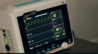
The head is strapped firmly so that movement does not hamper the imaging. The heart performance is observed by an ECG (Electrocardiogram), throughout the procedure. Then the catheter is injected and slowly passed through the arteries in the stomach, towards the neck and finally positioned for the entry of the dye material in the brain.
The dye material is inserted into the brain and the x-ray images are generated with a frame rate of 2-3 per second. Only expert radiologists operate this procedure. Slowly, every part of the brain artery network appears as black lines on a white background.
Once the imaging procedure is finished, the catheter is slowly withdrawn from the body. The point where the catheter was inserted is pressurized to stop bleeding. The leg cannot be moved and has to be kept straight, for up to 12 hours after the procedure is completed. Patients are usually kept under observation for sometime, to check for any abnormal reactions.
Angiogram Complications
Sometimes, the dye may flow out of the blood vessels. This indicates that there is internal bleeding in that part. The dye may get blocked in a vein, which indicates that there is a major blockage in that area. A misplaced artery may be an indication of brain tumor. It takes a trained and experienced doctor’s eye to detect the anomaly from a given image.
The brain is the most delicate part of the body and surgery of any part must be performed absolutely carefully. One mistake and the patient loses a body function, as brain is the controlling center. In this scenario, cerebral angiography is very important as when the surgeon knows exactly where the blood vessel anomaly lies, he can plan the surgery and perform it with minimum damage.
The procedure is the ‘roving eye’ of the neurologist, that has increased the success of brain surgeries and saved millions of lives, all over the world.


