Coronary angiography is used to examine blockages in the blood vessels. During the procedure, a special fluid is introduced into the coronary artery to detect the abnormalities that can cause heart diseases.
Coronary angiography is a method similar to X-ray imaging, and is used to examine the blood vessels and/or chambers of the heart. It is also referred to as cardiac angiography or coronary arteriography. It is performed in order to find out any blockage or narrowing of the blood vessels that may lead to heart attacks and other heart diseases. Usually, this procedure is recommended for those who have chest pain (angina), heart failure, and aortic stenosis.
In this technique, a catheter is introduced into a blood vessel, either in the upper arm or thigh portion, and the tip of the catheter is adjusted in the heart. A special dye is injected through the catheter that is detectable by X-ray. Certain precautionary measures are taken before the test. The patient needs to inform the physician if he/she has allergy problems or other underlying health conditions.
The Procedure
The patient is instructed not to eat or drink anything for 8 hours prior to the test. During the procedure, the patient lies down and is given a mild sedative for relaxation, but will remain awake during the procedure. Following this, an area in the upper arm or thigh is cleaned and numbed by administering local anesthesia. A small, flexible, hollow tube (catheter) is inserted into the blood vessel located in the numbed area. The other end of the catheter is moved gently along the blood vessel towards the aorta of the heart. While doing so, X-ray imaging is used to determine the path of the catheter.
Once the catheter is placed in the right coronary artery, a contrast dye is injected through the other end of the catheter, which is then circulated within the blood vessels. The patient may feel flushing of fluid in the blood vessels. Several X-ray images are taken following the dye injection. The recorded X-ray pictures are known as angiograms. These aer used for identifying any abnormal deposition, narrowing or enlargement of the blood vessels.
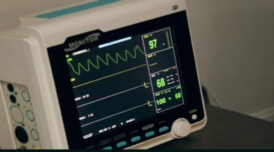
After recording the angiogram of a particular portion, the catheter is monitored in another major coronary artery. The procedure for injecting and recording is repeated. Several procedures are repeated in order to examine all the main coronary arteries and their branches. The whole test may last for one to several hours. During the procedure, the heart rate of the patient is monitored at all times.
When the test is completed, the catheter is pulled out gently. If the catheter insertion point is in the arm, the nurse may stitch the area and the patient will be able to move immediately after the procedure. In case, the insertion is in the thigh, then the patient should lie down for 10 – 15 minutes to prevent bleeding. The result of the coronary angiography is normal if there is absence of blockages and/or deposition in the blood vessels. A normal result shows proper circulation of blood to the heart. The result is abnormal in case of any blockages in the arterial walls. The angiogram is used for diagnosing the sites as well as the severity of blockages.
Coronary angiography is a safe procedure with minimal adverse effects. Some of the common side effects are bruise or infection at the insertion area, burning sensation, and mild angina during the test. Rarely, people with heart diseases could suffer from severe health complications such as a mild heart attack, damage to the artery, and at times, a stroke.


