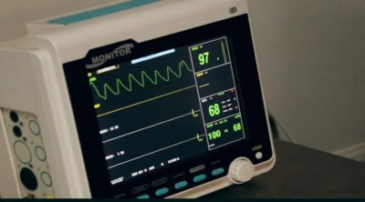
Electrocardiogram and echocardiogram are noninvasive procedures that check whether there are any issues with the heart. Here’s more about these procedures that evaluate heart health.
Did You Know?
The technology employed in an echocardiogram is identical to the one used in viewing a fetus during pregnancy.
Electrocardiogram (ECG or EKG) and echocardiogram (echo) are two important tests that investigate the heart’s functioning. Both tests provide an easy way to determine the health of your heart. These procedures can help diagnose an underlying abnormality in the working of the heart.
The following HealthHearty write-up discusses more on these procedures that examine the functioning of the heart.
Electrocardiogram vs. Echocardiogram
Working
✦ In an electrocardiogram, electrodes (small sensors) are attached to the chest, which measure the electrical activity of the heart. The electrodes are connected to a machine that plot a graph from the information it receives from the sensors. The graph plotted indicates the strength and pattern of electrical signals.
✦ Echo is done by keeping a small device (transducer) on the chest. The device generates high frequency sound waves that pass through the heart walls and are reflected back from the structures within the heart. The transducer receives these returning sound waves, which are then processed by a computer to generate a 2D moving image of the heart.
Function
✦ The ECG test checks your heart’s electrical system. It is this system that generates electrical signals that convey the heart when to beat. These tiny electrical pulses that regulate heartbeats are recorded by the ECG device. The test also measures the signal strength and verifies whether the heart is sending steady electrical signals.
An ECG also records the timing of your electrical signals, which indicates how fast your heart is beating. It evaluates the basic rhythm of the heart, and any disturbances in it can be easily recorded by the ECG.
✦ An echocardiogram can check the pumping capacity of your heart. It determines how well your heart pumps blood around the body, when it beats. It can check whether the heart’s pumping action is becoming weaker, a symptom associated with heart failure. It can calculate the amount of the blood that is pumped out with every heartbeat, thus, indicating the heart’s pumping strength.
Output or Results
✦ As aforementioned, the output of the ECG device is a graph that indicates the intensity and direction of electrical signals generated in the heart. The heart function is evaluated depending upon how the electrical signals get traced on the graph.
✦ With an echo, you see the live pictures of the insides of your heart. The echo test shows a 2D cone-shaped real-time motion of the heart on a monitor. Experts can actually view how the heart is beating. As a result, they are in a better position to evaluate the functioning of the heart. With advancements in the medical field, now an echo test can also show a moving 3D image of the heart. However, it is usually used to diagnose some rare complications of the heart.
Issues that Can Be Detected
✦ An ECG test can help diagnose heart rhythm problems such as heart arrhythmia that causes irregular heartbeats. It can detect if the heart is beating too hard (tachycardia) or too slowly (bradycardia). These issues often arise when the heart’s electrical activity is not working properly.
✦ A moving 2D image generated by an echo test allows accurate assessment of the heart valves. There are 4 valves, each one ensuring one-directional blood circulation through the heart. Heart valve disorders such as stenosis, atresia, and regurgitation that disturb the blood flow through the heart can be detected using echo. It can also determine how severe the valve disease is.
An echo scan can also detect sections of the heart muscle that are not working properly. It can also identify the causes of poor heart muscle function, such as inadequate blood circulation from an earlier heart attack. An echo can also help assess the heart function after a heart attack. Possible blood clots within the heart can also be located using the test.
Similarity
✦ Both ECG and echo tests can evaluate the size of the chambers of the heart. For instance, heart conditions, such as left or right atrial enlargement, can be detected through an ECG test. These tests can also identify abnormal positioning of the heart. Other cardiac structural abnormalities, such as myocarditis (swelling of the heart muscle) and pericarditis (inflammation of the tissue surrounding the heart), can also be diagnosed using an ECG and echo.
Why an Echocardiogram is Better
✦ As an echo scan provides accurate moving 2D visuals of different structures within the heart, the test is more reliable at judging heart health and its pumping action as opposed to an ECG. The direct visualization of the heart chambers in an echo scan is what makes the procedure more accurate in diagnosing heart ailments.
✦ The clarity with which the results are obtained through an echo is something that one cannot expect from an ECG test. Structural abnormalities, such as thickened heart muscle, that need to be interpreted on an ECG, are distinctly visible on an echo test. However, an ECG is better at detecting abnormal heart rhythms that cause heart palpitations, but even then, the doctor may recommend an echo rest to find out the cause of heart rhythm irregularities. On the whole, though an ECG can detect abnormalities in the heart structure, a confirmation is often necessary through an echocardiogram.


