Neuroblastoma is cancer of the nerve tissue and observed in infants and young children, usually under the age of 5. There are several ways to detect the presence of neuroblastoma in a child. Here is a summary of different methods to diagnose neuroblastoma.
Neuroblastoma refers to malignancy in the nervous tissue, specifically the sympathetic nervous system. The sympathetic nervous system is concerned with certain body functions such as heart rate and blood pressure. According to PubMed Health, about one in 100,000 children is affected by cancer of the nerve cells.
Neuroblastoma goes undetected partly due to the fact that the symptoms become evident after the tumor has metastasized to several parts of the body. This is also because this nerve cell cancer occurs in babies, and its symptoms mimic common illnesses and viruses. Besides infants may not be able to express the pain and symptoms experienced clearly and accurately. The exact causes cannot be pin-pointed. Etiology suspects contribution by parental factors such as consuming alcohol, smoking, consuming medicinal drugs during pregnancy and childbirth. However, a genetic factor has also been elicit; familial cases have been identified.
Diagnosis of Neuroblastoma
The symptoms of neuroblastoma become evident only after the tumor has metastasized to different parts of the body. This may make one wonder how this condition is diagnosed. Several changes have already taken place in the body by the time the patient turns up for diagnostic testing; so this cancer can be detected and confirmed at various levels – organs, tissues, blood samples, etc. On observing likely symptoms, a doctor may suggest one or more of the following diagnostic tests. The rationale behind diagnosing neuroblastoma is –
- General physical examination (whether the tumor is suspected to be present in one or more body parts depends on the degree of metastasis that has occurred)
- Primary tumor is located to detect metastasis pattern
- Additional tests may be carried out for further study and to devise treatment
General Physical Examination Tests
Palpation of Abdomen: The examiner will feel different parts of the patient’s body with his/her hands and fingers, and thus verify the size, consistency, tenderness of different body organs.
Rationale Most neuroblastomas initiate in the abdomen, hence it is the first body part to be checked.
Observation Unusual responses (if any) of patient to palpation are noted.
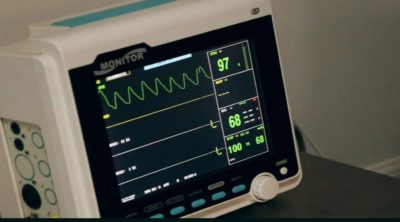
Blood Pressure and Pulse: On an average, normal blood pressure of an infant is: systolic > 90 mmHg and diastolic > 60 mmHg
Rationale If a tumor metastasizes to the adrenal gland, it most significantly affects the blood pressure and heart rate of a patient.
Observation Patient most commonly shows elevated blood pressure and heart rate.
Swollen Lymph Nodes: Physical examination of the limbs can detect swollen lymph nodes.
Rationale Metastasis of neuroblastoma leads to swelling of the lymph nodes.
Observation Lymph nodes appear swollen.
Tests For Locating the Primary Tumor
*Results are confirmed by carrying out more than a single test
Bone Scan: A bone scan involves the injection of a minute amount of radioactive material in the vein. As it courses through the body, it will locate in different body parts and will give off radiation, which is detected by a camera.
Rationale If a tumor is located in the bone, the bone turnover (or metabolism) will increase, hence more radioactive material will locate there (than normally does) and hence more radiation will be emitted.
Observation Detection of radiation “hot spots” in a bone scan suggests the presence of a tumor.
X-ray and CT Scan: Usually an x-ray of the bones and chest, and a CT scan of the chest and abdomen is performed. A CT scan enables one to view internal body parts without superimposition. CT stands for computed tomography.
Rationale The x-ray reveals the presence of tumors. a CT scan will reveal tumors that remain concealed in an x-ray (since a CT scan will generate images without the effect of superimposition).
Observation Presence of tumor, and hence stage of neuroblastoma can be estimated by such imaging.
MRI Scan of Chest and Abdomen: Procedure of MRI (Magnetic Resonance Imaging) scan involves applying a magnetic field to the patient’s body (or a certain body part). The magnetic field causes nuclei of atoms in the patient’s body to ‘align’ and ‘resonate’ in a particular fashion. This resonance is detected and used to construct an internal image of the body part being scanned.
Rationale The presence of tumor may lead an organ to emit unusual resonance, and thus distort the constructed image.
Observation A distorted image indicates the presence of tumor in the scanned body part.
Additional Tests
*These tests are performed as an additional confirmatory level.
Bone Marrow Biopsy: A small amount of bone marrow is aspirated using a needle from the hip bone and then microscopically observed.
Rationale In case the tumor has metastasized to the bone, abnormal proportions of different cells may be seen in the marrow. The morphology of the cells may also change.
Observation Presence of tumors can be detected. Nature of tumor (benign or malignant) can also be determined.
CBC (Complete Blood Count): CBC involves enumerating the number of cells of each blood cell type to detect abnormal proportions.
Rationale One of the complication of neuroblastoma is anemia.
Observation Abnormally low proportions of red blood cells indicates anemic condition.
mIBG scintiscan: mIBG (metaiodobenzylguanidine) scintiscan involves injecting radioactive mIBG molecules into the vein. The sympathetic neurons take up this molecule.
Rationale mIBG is a functional analog of norepinephrine. Norepinephrine is a neurotransmitter and is taken up by sympathetic neurons. Since mIBG molecules are similar in structure and function to norepinephrine, they are also taken up by sympathetic neurons. A tumor in the sympathetic neurons (as observed in neuroblastoma) will hence cause more mIBG molecules to be taken up.
Observation A radiation “hot spot” detects the presence of tumor in sympathetic neurons, i.e. neuroblastoma.
Urine Test: The urine of the patient is examined for elevated levels of catecholamines, dopamine, HVA (homovanillic acid), VMA (vanillylmandelic acid).
Rationale Nerve tissues of the body (including sympathetic neurons) synthesize catecholamines and related metabolites in the body. Presence of tumor will increase the amounts of these metabolites synthesized in the body.
Observation Elevated levels of catecholamines and related metabolites indicates neuroblastoma.
Diagnosis of any disease (including neuroblastoma) cannot and does not depend on a single test. Often multiple tests are carried out to confirm the results obtained in preliminary tests. Only if all the test results conform, the patient is declared to have or not have a particular disease. Doctors and health care providers are more than willing to answer all doubts and queries related to the patient’s ailment and troubles. The patient’s parents should take efforts to understand test procedures thoroughly and should educate themselves as much as they can. That way they can prepare their children for the tests better.
Disclaimer: This article is purely for informative and educational purposes and does not advice. If users need medical advice, they should consult a doctor or other appropriate medical professionals.


