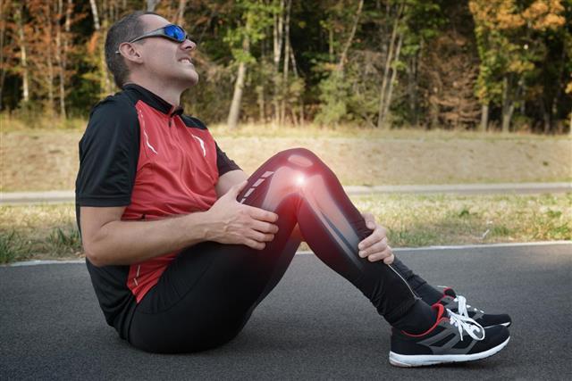
Medial meniscus, situated in the knee joint plays the role of a load bearer, shock absorber, knee stabilizer, etc. Injury to the medial meniscus is called medial meniscus tear.
Medial meniscus tears are common knee injuries and occur in about 61 per 100,000 people. Surgeries pertaining to meniscus are carried out on approximately 85,000 patients each year. Then how is it that we don’t hear this term being used so often? The reason being medial meniscus tear is most commonly known as ‘torn cartilage’. Athletes are often seen to suffer from this kind of an injury, as they often encounter traumatic forces while playing or training for the sport.
Anatomy of Meniscus
The knee joint – the largest joint in the body is a complex joint and consists of three bones: Femur bone (thigh bone), Tibia (bone of the lower leg) and the Patella (knee cap). Each of these bones are covered by a thin layer of articular cartilage, which is hard but smooth in nature and reduces friction between the bones. Besides the bones, four ligaments are also present: Anterior cruciate ligament (ACL), Posterior cruciate ligament (PCL), Medial collateral ligament (MCL) and the Lateral collateral ligament (LCL) which stabilize the knee, thereby providing strength to the knee joint.
There exists two wedge-shaped, thick, rubbery menisci (plural of meniscus) situated in each knee, that are located on the medial (inner side) and lateral (outer) sides of the knee joint. Based on their location they are termed as medial meniscus (C- shaped) and lateral meniscus (O-shaped). Medial meniscus is slightly larger and is only half as mobile as the lateral meniscus. The peripheral, convex border of the meniscus is thicker as compared to the inner/central portion.
The menisci, sandwiched between the femur and the tibia are held in place by the coronary ligaments. The wedged shape of the meniscus enables it to keep the rounded condyles of the femur from sliding off the almost flat tibial surface. The meniscus is popularly known as the ‘cartilage’ of the knee, however, it is composed of dense fibro-cartilage, making it flexible and not as hard as articular cartilage.
Small blood vessels provide blood supply to the meniscus. However, not the entire portion of the meniscus receives blood supply. The central portion of the meniscus is avascular (no blood supply). The menisci are divided into various zones based on the blood supply available to them, namely, the red zone (peripheral meniscus) which receives vascular (blood) supply, the red-white zone with blood supply only in its outer region and the white zone (central meniscus) which lacks blood supply.
Medial Meniscus
The C-shaped medial meniscus is asymmetric, with the anterior horn wider than the posterior horn. It is approximately 3.5 cm in length and is attached to the medial collateral ligament (MCL). The peripheral part is attached to the joint capsule and the middle portion is firmly attached to fibers of the medial collateral ligament. Thus, it is common to find medial meniscus tear occurring with simultaneous tears in the anterior cruciate ligament and the medial collateral ligament due to their interconnections. This triple rupture phenomenon is called ‘unhappy triad’.
Function of the Meniscus
Shock Absorber: The menisci, cushion the forces which are directed upward or downward through the knee. They spread out the forces being transferred from the thigh to the lower leg and diminish the load borne by the spine, pelvis, hip, ankle and foot joints.
Protects Articular Cartilage: The menisci prevent the wear and tear of the articular cartilage by spreading out the forces (during walking, running) applied on the knee joint surfaces. In the absence of the meniscus, a large amount of force would have been concentrated on the small area of the articular cartilage leading to damage and degeneration (osteoarthritis).
Nutrition: The repeated loading and unloading action of the joint helps in lubrication and nourishment of the cartilage. The menisci also assist in nutrition of the knee joint.
Knee Stability: The wedge shape of the meniscus helps it to deepen the almost flat tibial surface into a shallow socket, thereby allowing the femur to easily slide over the tibia.
Even distribution of body weight: The meniscus evenly distributes the forces applied on the knee joint, by distributing the forces across a large area and minimizing focal contact pressure.
Load bearing: The meniscus bears around 40% of the total weight applied on the knees and reduces the load on the articular cartilage.
Medial Meniscus Tear
Twisting or application of abnormal pressure on the medial meniscus causes the meniscus to get jammed between the bones, thereby resulting into tearing or splitting of itself and is called medial meniscus tear. Medial meniscus gets 10 times more frequently injured, as compared to the lateral meniscus. This is because the medial meniscus is more firmly attached to the tibial surface and knee joint capsule.
Traumatic or stressful activities encountered during sports (football, rugby) involving sudden twisting of the knee when the foot is planted on the ground (weight-bearing position) can result in medial meniscus tear. Most commonly, such tears are seen to occur in situations when the knee is in the bent position and twisting occurs on the bent knee. Severe pain and swelling within 24 hours occur subsequently.
Loss of resiliency of the meniscus can also lead to medial meniscus tears. This is seen in older people whose meniscus happen to degenerate on aging and is termed as ‘degenerative tear’. Sometimes, when the tear is mild, it goes unnoticed. However, years later it may resurface by a movement as mere as walking, tripping or squatting. Injury to the medial meniscus can happen to people of all ages. However, the healing process in every age group is different. In younger people, the meniscus is tough and rubbery, whereas in older age groups the meniscus becomes weak with age. Young people’s menisci respond to treatment more readily as compared to older people, who have unfavorable outcomes even after surgery.
The size and variety of each medial meniscus tear can vary widely, thus the severity of the tear depends on the size and type of tear. The different types of medial meniscus tears are bucket handle tears (longitudinal tear with the shape of a bucket handle), radial tears, horizontal tears, parrot beak tears and complex tears. The medial meniscus may be torn in half, cut off in the shape of a C along the circumference or can also hang loosely from the knee joint by a thread. The zone of meniscus where injury occurs also matters. If the injury has occurred at the peripheral region, then healing will take place faster, due to the blood supply. However, for injuries occurring at the white zone, the lack of blood supply hinders the healing process.
Symptoms of Medial Meniscus Tear:
- Pain in the knee area
- Swelling of the knee joint
- Increased temperature of the knee, inflammation
- Stiffness and tightening of the knee
- Flexing is possible, however leg extension becomes painful.
- Difficulty bending, squatting
- Walking becomes difficult
- Instability and performing normal activities become difficult.
- On pressing the knee joint line, tenderness is produced.
- Sensation that the knee is giving away
- Knee clicking
- Knee catching
- Knee locking
Knee locking is a condition in which one cannot extend the knee. This happens when a loose piece of the meniscus gets stuck in the knee joint causing inability (temporary) to extend the leg completely. Only larger and more severe tears result into knee locking and giving way signs.
Diagnosis and Treatment
People with a meniscus tear will know there is a problem due to the pain emanated and swelling followed in a few hours time. Some may even hear a pop in the knee. When the meniscus pain aggravates, the person will know something is wrong. This is when the early treatment comes into place.
Early Injury Treatment: The ‘RICE’ treatment is followed to control the swelling and pain, for the first 72 hours after the injury. RICE stands for Rest, Ice, Compression and Elevation. The wounded person must restrain from any kind of pain aggravating activities. An ice pack is placed for the next 72 hours, which must be applied 15-20 minutes at a time. The ice however is not allowed to come in direct contact with the skin and is placed in a wet towel or cloth. Elastic bandage is used to compress the injured area. The fourth step is to elevate the injured portion above the heart level, so as to reduce the swelling.
Scan and X-rays: From the meniscus tear symptoms, a medical practitioner can diagnose meniscus injury via physical examination. He will check the mobility, swelling and tenderness in the knee joint. However, he will still need to get diagnostic imaging done in order to determine the extent of the injury. The X-rays usually do not reveal medial meniscus tears. Magnetic Resonance Imaging (MRI) is used for meniscus tear diagnosis. The meniscus normally appears black on the MRI, while the tears will appear in the form of white streaks in the MRI, which helps diagnose the tear. Depending on the results of the diagnosis of the tear, treatment will be carried out. A knee arthroscopy may also be carried out to detect the specific portion of the tear and its severity. However, because MRI clearly shows almost every joint in the body, most surgeons prefer MRI over arthroscopy. When the MRI results are not clear, arthroscopic method is used, which is also less expensive, less painful and involves lesser recovery time.
Conservative/ Traditional Method: Small tears at the outer edge of the meniscus can be repaired without surgical intervention. For some fortunate folks, the medial meniscus rebuilds itself, by taking rest, application of ice packs, physical therapy and ingestion of non-steroidal anti-inflammatory medicines. Patients undergoing this traditional form of treatment have to refrain from performing routine activities like running, jumping, etc. Taking complete rest and allowing the knee to heal is mandatory.Rehabilitation knee exercises are carried out later to strengthen the muscles around the knee, under the guidance of a physical therapist.
Surgery: Cases wherein physical therapy fails to bring about healing and where knee locking has occurred, surgical intervention is required. Depending on the severity of the injury, the surgeon may either repair, remove or replace the meniscus. Let’s have a look at all three.
•Knee Arthoscopy
The tear can be repaired by arthroscopic method. In this method, two small incisions are made in the knee joint through which the tiny camera is sent, in order to enable the surgeon to get a full view of the internal parts. Then the surgeon inserts tiny instruments into the knee joints and conducts the repair surgery. Once the surgery procedure ceases, a knee cast is placed on the knee externally to prevent mobility and facilitate healing. After initial progress, the surgeon will recommend specific knee strengthening exercises. The type of surgery reduces the amount of damage that can otherwise be done during surgery, is less painful and promotes more fuller and speedy recovery.
•Partial Meniscectomy
In cases where the meniscus tear is too severe and beyond repair, the torn portion of the meniscus will have to be surgically removed. This is because the torn portion ceases to function as the normal meniscus and is of no value, moreover, leaving it in the knee will only conduce to irritation. Thus, it is wise to remove the torn part of the meniscus, before it causes a larger tear. This procedure is called partial meniscectomy and is done arthroscopically. The torn portion is discarded, while the rest of the meniscus is left intact after smoothening any loose ends.
•Total Meniscectomy
In this case the whole meniscus is surgically removed. However, removal of meniscus results in various long-term debilitating effects on the knee. Which is why meniscus transplant is carried out to replace the lost meniscus tissue using donor tissue. Donor graft (taken from a cadaver) is tested for diseases, sterilized and sized in the lab, according to the size of the patient’s tibia. The re-sized donor tissue is then stitched into the gap where the original meniscus was positioned. Meniscus tear recovery time for this surgery will vary from anywhere between 4-8 months.
A severe medial meniscus tear can bring about drastic changes in a person’s life. Performing daily routine activities, and work and play become difficult. Thus proper functioning of the meniscus is crucial for the health of not only our knees, but also our lives.


