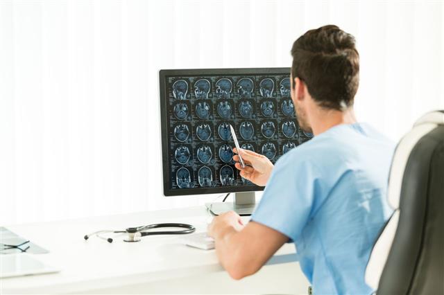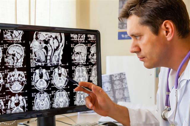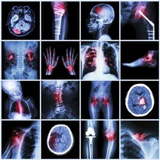
A CT scan with contrast uses certain substances that are given orally or intravenously before the scanning procedure, in order to improve visibility of resultant images.
A CT scan is a technique that gives detailed images of the inside of the body. This is a painless procedure that combines the power of a sophisticated X-ray machine with that of a computer, to generate a 3D view of the inside of the body. This advanced method provides an easy way to inspect any organ of the body in much finer detail. In this procedure, the X-ray equipment rotates around the body and sends several beams to take multiple pictures of the same area from different angles, which are then put together by the computer to reveal an extremely detailed cross-sectional 3D image of the body part under examination. A normal X-ray machine uses only one radiation beam to produce an image that is not capable of giving a clear idea about the disease.
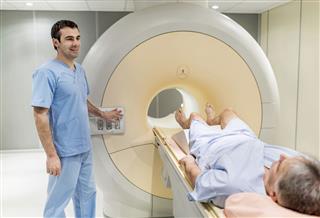
Before the procedure actually begins, you will be advised to lie down on a rectangular table. The table is then pushed until you come exactly at the center of the CT scanner machine. During the procedure, the CT scanner is seen revolving around you, clicking images from different angles. While the imaging is going on, one should not move as the resultant images appear distorted.
CT Scan Benefits
- CT scans have been extremely useful to diagnose diseases and disorders in the early stages of development. Doctors find this procedure beneficial to detect tumors early, which in turn helps to plan the treatment to prevent further complications.
- CT scanning could determine whether an artery wall has swollen, as well as find out the severity of internal injuries.
- CT scanning can also be used to know how the body is responding to treatments, like radiation therapy and chemotherapy.
- In order to diagnose diseases related to bones, such as osteoporosis, the bone mineral density needs to be ascertained, which can be effectively done using a CT scan.
CT Scan With Contrast
No doubt, CT scanning depicts sharp images of different organs but when it comes to evaluating small structures of the body, better visibility is necessary, which unfortunately CT scan alone does not provide. CT scanning that is done with the help of contrast agents can play a significant role to improve the images of the area to be examined. For better visualization of a specific area within an organ, a CT scan with contrast is a good option. Detecting inflammation in a specific part of an organ due to an infection may not be possible in the absence of contrasting agents. Unless the portion is highlighted, diagnosing it is not easy. For instance, CT scan of gastrointestinal tract shows the stomach, small and the large intestine. But, scanning with a contrasting agent will allow doctors to differentiate these organs clearly.
Contrasting agents enhance the visualization, which helps to correctly diagnose the condition. So, when it comes to making the structure of interest more visible, what better way than CT scan with contrast. This method provides superior images that allows doctors to get a better assessment of the condition. The contrast, also known as a dye, is basically a substance that is administered intravenously or taken orally during the CT scan test. These substances have the capability to weaken (attenuate) X-rays. When these substances are taken, they travel in the body and are absorbed by the organs. When the radiation beams pass through the organ containing the contrast, these X-rays attenuate significantly. As a result, the organs or the tissues that contain the contrast are highlighted, which appear as white areas on the final CT images.
- This method that involves use of contrasting agents increases the visibility of the organ to be studied. For diagnosing tumors, and to know the exact location of the tumor, sometimes a CT scan with contrast is used.
- A blood clot deep into the venous system, or blood vessel defects can be easily detected with this procedure. In other words, to find out blockages in tiny blood vessels, use of CT scan with contrast is necessary.
- This new innovative technique enhances particular structures of the organ which helps to identify the abnormalities in the organ.
- Patients undergoing this type of CT scan are instructed not to consume solid food for at least 4-6 hours before the test.
- However, the patient is allowed to drink clear liquids such as water, apple juice, tea, or clear soda, prior to the CT scan.
Contrast Agents
During a CT scan, the patient may be advised to take a contrast (dye) so that resultant images of a specific part of the body have greater clarity. This may be essential to detect the presence of a foreign object, or to diagnose any minute defects in a specific organ. When the image of a specific body part is produced using a contrast agent, certain areas of the image are highlighted. In other words, a precise visualization of the specific area of the organ is achieved, which in turn helps to asses complications in the organ. Some of the most commonly used contrasts are barium, iodine, barium sulfate, and gastrografin. Contrasts are mainly administered in 4 different ways which are listed below:
- Intravenous (injected through a vein to highlight blood vessels and to get a clearer image of the structure of the organs such as the spine, liver, and the brain)
- Oral (taken by mouth for an abdomen CT scan)
- Rectal contrasts to highlight large intestines
- Inhalation
Administration of contrast agents by allowing the patient to inhale a gas, namely xenon, is an uncommon method and is followed when highly sharp images of the lungs or the brain are required. Also, this procedure is conducted at very few locations worldwide and only in rare cases.
In most cases, solution laced with diluted barium (that acts as an oral contrast ) is given to the patient before undergoing abdominal CT scan. Around 1/2 to 1 liter of oral contrast has to be consumed in a span of two hours. However, those receiving contrast intravenously (through injection) should avoid consuming both solid and liquid products as there is a high probability of suffering from stomach upset.
Chest CT/Contrast
A chest CT scan using contrast gives detailed images taken at varying angles of the different parts of the chest. The contrast media is generally given intravenously. It is a non-invasive procedure that provides increased visibility of the structures of the chest that include the lungs, heart, and surrounding bones. This method gives precise images that help to determine the presence and extent of injury. Chest CT scanning with contrast gives an in-depth view of inside the chest. This multiple-angled X-ray technique takes many sharp images of the chest.
- People having chest pain or symptoms of lung disease or difficulty in breathing may require chest CT scans with contrast.
- The procedure helps to diagnose the root cause of the problem. It looks for abnormalities such as lung tumors, blood clots, or excess accumulation of fluid around the lungs. Even conditions like pneumonia and tuberculosis can be detected with this method.
- Patients advised to undergo chest CT scans need to wear loose-fitting clothing and remove items like jewelry and other metal objects as they may interfere with the working of the machine.
Abdominal CT Contrast
In order to scan the abdomen and the large intestines, rectal CT contrasts are often administered using an enema. The most commonly used rectal contrast are gastrografin and barium sulfate, which help to enhance the images of the large intestines as well the bladder, uterus (womb) in women, and other organs contained in the pelvis. When gastrografin as a contrast is used, gastrointestinal organs that are within the abdomen are highlighted clearly in the resultant image. Sometimes, gastrografin, available as a water-based drink, is consumed to about 1000cc or 1500cc 1-2 hours prior to CT scan to get the desired result. Pregnant women are advised to stay away from abdominal scans as exposing the fetus to several beams of radiation can cause health complications in the unborn child.
Head CT Scan with Contrast
Head CT scan is often used to detect abnormalities in the brain such as tumors. The scan can help to diagnose a stroke by pointing out blood circulation problems in a specific part of the brain. It can pinpoint the arteries where blood flow has been obstructed. A head CT scan is also used to investigate jaws, sinuses (the hollow air-filled spaces around the nose) for infections such as sinusitis. People suffering from frequent dizziness, chronic headaches, or neurological disorders such as seizures may also be advised to undergo a CT head scan.
One cannot wear hearing aids, dentures, braces, and other such metal objects during a head CT scan as they can distort the resultant images. Barrettes (hair clips), eye glasses, and contact lenses are also not allowed in the CT scanner.
CT Scan Duration
The amount of time that you will spend lying still during the CT scan depends on the size of area under investigation. Larger the area of CT scan, more will be the duration of examination. Also, scanning that involves use of contrast agents is more time-consuming. In general, the CT scan examination takes a minimum of 15 minutes but the duration can extend up to 60 minutes.
Results
Although the images are generated in seconds and the procedure lasts about an hour, you do not get the results immediately nor in a couple of days. The radiologist studies and interprets the images and finally conveys it to your health care provider. So, it may take a couple of weeks before the doctor gives you the CT scan report.
Side Effects
Contrast agents that are either given orally or intravenously are eliminated out of the body through urine. So, as such bothersome side effects are unlikely to occur. However, serious health complications will occur if the patient is allergic to the contrast dye. After contrast is administered intravenously, you may experience a warm feeling that usually goes away in a short while. It is observed that oral contrasts have a bitter taste that lasts for a few minutes. In general, the side effects that may occur by using this type of CT scan are listed below:
- Vomiting and nausea
- Chills
- Fatigue
- Sneezing
- Dizziness
- Flushing
- Metallic taste in mouth
- Skin problems such as hives
- Constipation, in case of rectal contrasts
- Allergic reactions, such as swelling of the throat and difficulty in breathing
In case the patient is allergic to contrast agents, the doctor should be immediately informed so that he can take precautionary measures to prevent any adverse reactions.
CT Scan With Contrast Risks
Kidney Damage
It is crucial that people recommended for CT scan with contrast are not suffering from kidney diseases. The contrast agents that are administered either intravenously or orally will be flushed off through the urine, only if the kidneys are working correctly. The iodine contrast can cause serious damage in people with compromised kidney function. So, a blood test that evaluates kidney function is mandatory, 3 days prior to CT scan with contrast. In case, the test indicates impaired kidney function, use of contrast agents is avoided.
Cancer
A CT scan with contrast exposes you to radiation that is significantly more in magnitude than that of conventional X rays. So, frequent exposure to CT scan can put you in the risk zone of cancer. Attending just one session of CT scan also carries a small amount of cancer risk. Although high radiation exposure from CT scan is not a major risk factor in the development of cancer, unnecessary usage of this radiation-based procedure needs to be avoided. CT scan generates radiation that is 100 to 500 times more intense than the radiation delivered by the X-rays. Exposure to such high-intensity rays may inflict damage to DNA and cause cancer in the long run. So, to be on the safer side, excessive use of CT scan has to be curbed.
Protecting Kidneys from Contrast Dye
A study published in the Annals of Internal Medicine showed that taking a N-acetylcysteine tablet before a CT scan can substantially reduce the risk of kidney damage resulting from iodine contrast agents. As aforementioned, exposure to intravenous contrast dye can lead to kidney problems. This is also true for people with healthy kidney function, although the risk is minimal. However, taking this medication can help to keep the kidneys safe from contrast-induced nephropathy.
CT Scan Vs. MRI Scan
A CT scan is not the same as an MRI scan. This is because an MRI scan uses magnetic fields to generate images of internal organs as compared to X-ray beams used in CT scans. Also, MRI scans are used only in a few situations, like diagnosing brain tumors and primary bone tumors. CT scans, on the other hand, still remain one of the best medical tests and tools for early detection of various diseases.
Disclaimer: The information provided in this article is solely for educating the reader. It is not intended to be a substitute for the advice of a medical expert.
